Scientists make organs transparent so you can see inside
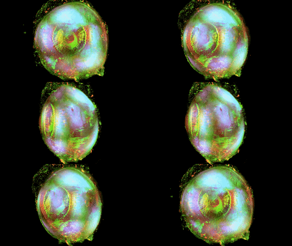
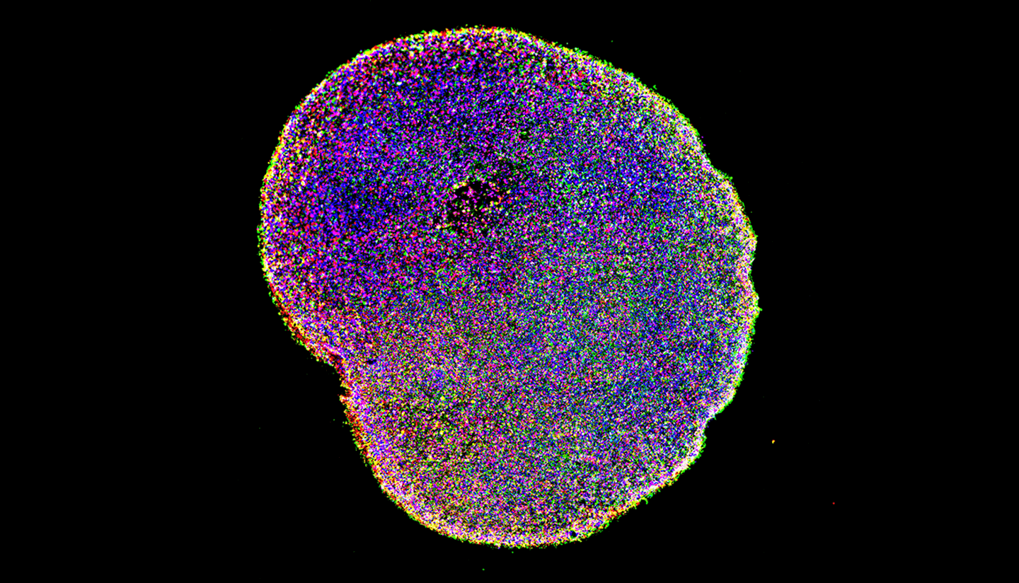
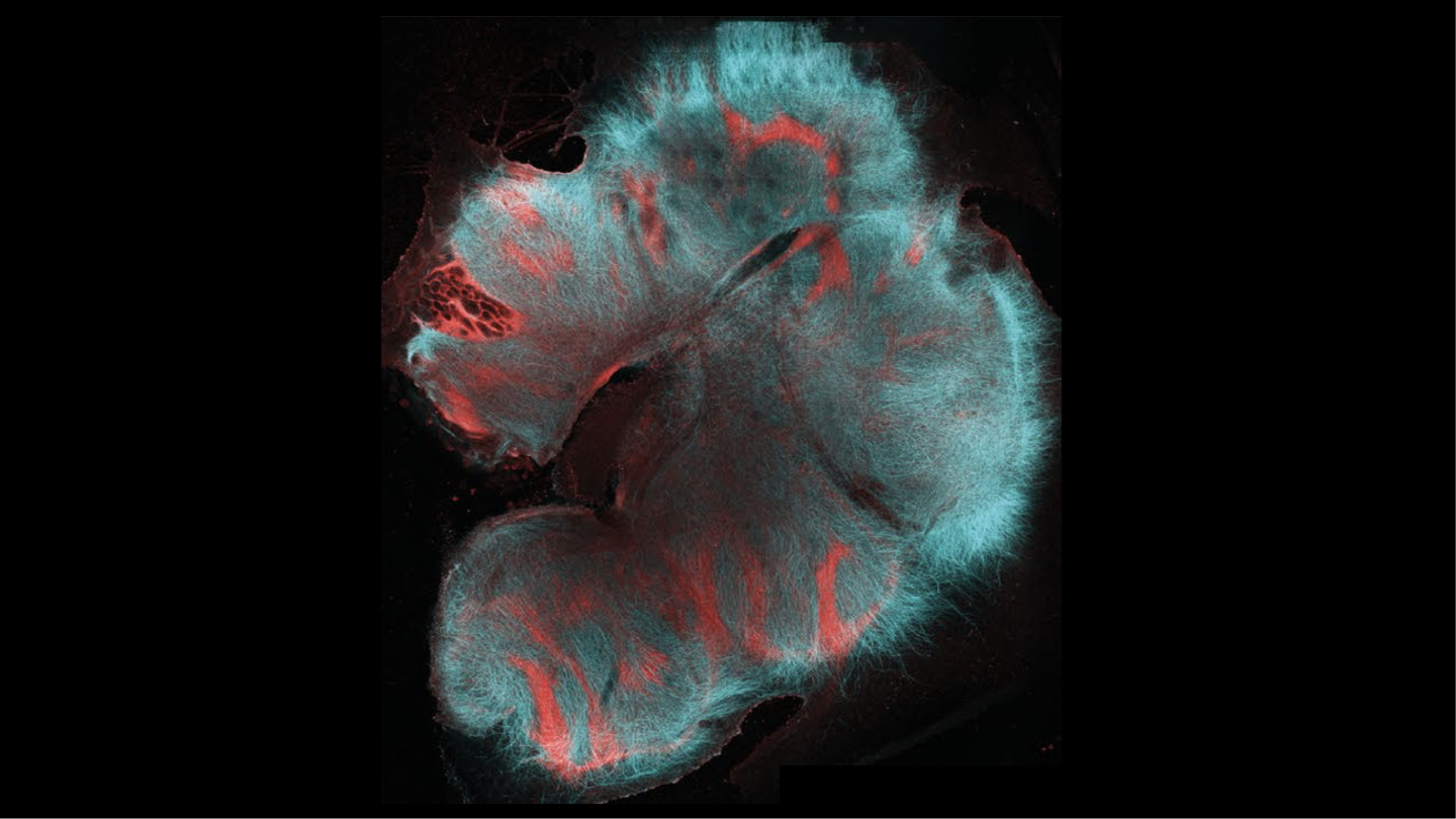
Image source: Zhao, et al
- By manipulating light refraction in organ tissue, it can be made transparent.
- Coloring internal structures is as “simple” as slipping dyes between tissue cells.
- A new method paves the way for fully 3D imagery of mature human organs.
As science dives deeper into the physiology of human organs, and in particular the human brain, it has become clear that viewing such organs in three dimensions and in microscopic detail is of critical importance if we’re ever to understand how they work. Of course, organs are solid, and seeing what’s going on inside them has proven a challenge to say the least. Now a new system, the SHANEL method, has been announced. It provides a practical solution for doing just this, and the glimpses it gives us inside the human brain and other harvested organs are stunning.
All of the videos below are by Zhao, et al.
Making tissue transparent
SHANEL addresses two problems. The first is transparency: You can’t simply look through the tissue encasing and surrounding the structures we’re interested in observing.
Scientists have attempted for some time to explore the brain by slicing it into thin sections. However, reassembling such sections into a 3D model can take years, and the final assemblage is likely to exhibit distortions introduced by the tissue-slicing process.
In the last 10 years or so, scientists have been refining a process called “optical clearing.” Its goal is not to remove all the tissue hiding organs’ secrets, but to make it transparent.
What causes tissue to be translucent, that is, not transparent, is light scattering. Tissue is made primarily of water surrounded by lipids and proteins, all of which refract light to different degrees. Using a refractive index (RI), for example, water has an RI of 1.33, proteins more than 1.44, and lipids more than 1.45. Together, light bouncing around these components render the tissue translucent.
Various optical cleaning methods have been developed that remove, modify, or replace tissue components using either solvents or water-based methods to normalize their RI values and “clear” the tissue in preparation for microscopy.
While progress has been made in clearing rodent organs and human embryos, say the developers of SHANEL, “Adult human organs are particularly challenging for this approach, owing to the accumulation of dense and sturdy molecules in decades-aged human tissues.” In their announcement they report a case in which clearing even an 8 mm-thick slice of human brain took 10 months to clear, and another where 3.5 months were required to clear just a 5 mm-thick human striatum sample. Clearing entire organs or larger segments of them has been out of the question.
SHANEL stands for “Small-micelle-mediated Human orgAN Efficient clearing and Labeling,” (we’ll get to the labeling in a moment.) It uses a new, small micelle — a detergent aggregate of surfactant molecules in a liquid colloid — that can permeate centimeters-thick mammalian organs and clear them. A key ingredient of it is the zwitterionic detergent known as CHAPS. CHAPS’ unusual chemistry contains a structure of both hydrophobic and hydrophilic faces, resulting in much smaller micelles that can permeate tissues more effectively than other detergents.
Molecular-level labeling
That final letter in SHANEL, as noted, stands for “Labeling,” which is another problem that’s vexed scientists and for which SHANEL provides a new solution. While seeing inside organs is a large part of the battle, another is developing a means of marking molecular structures so they can be differentiated from other components of the organs. This is generally done using a method called “permeabilization,” that sneaks dyes and such between tissue cells to mark the desired objects. SHANEL’s smaller, micelles greatly improves scientists’ ability to reach and make desired targets visible in imagery, as the videos here show so dramatically.
Just the beginning
For now, SHANEL’s inventors assert they can clear “human samples ranging from 1.5 cm thickness to whole adult human organs,” adding, “We also show that the technology works on other large mammalian organs such as pig brain and pancreas.” Going forward they expect to “pave the way for cellular and molecular mapping of whole adult human organs, including the human brain.”
We imagine you agree they’re already creating some pretty amazing imagery.
