A man-made embryo shows how a stem cell finds its role
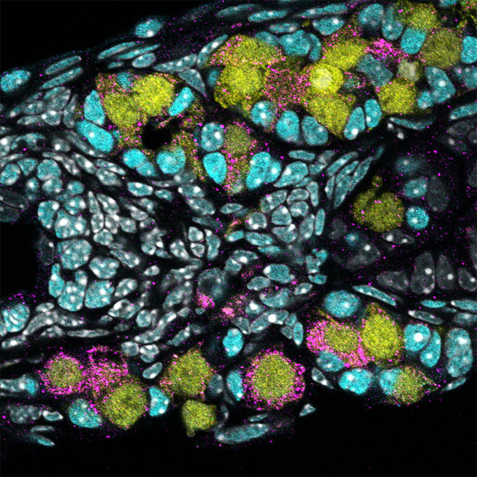
A unique 3D model allows researchers to explore embryonic development.
- Researchers observe the beginning of embryonic stem cells dividing into upper and lower body sections.
- An interdisciplinary team invents an impressively accurate 10-day-old "embryoid."
- The team's model may be important to other future research on pregnancy.
Breaking symmetry
<img type="lazy-image" data-runner-src="https://assets.rebelmouse.io/eyJhbGciOiJIUzI1NiIsInR5cCI6IkpXVCJ9.eyJpbWFnZSI6Imh0dHBzOi8vYXNzZXRzLnJibC5tcy8xOTcwODI1MS9vcmlnaW4uanBnIiwiZXhwaXJlc19hdCI6MTY3NDk3NDk3NH0.V9iZeyWqNwWru5QqQkd6mtl28ZoCqOidYUzq1to4juc/img.jpg?width=980" id="0988c" class="rm-shortcode" data-rm-shortcode-id="bb4064f8b3064071e609d5687496756b" data-rm-shortcode-name="rebelmouse-image" />Artist's rendering of stem cells
Image source: Giovanni Cancemi/Shutterstock
<p>Earlier research using mouse embryoids demonstrated symmetry breaking, and it was observed in human embryonic stem cells a few years ago, leading to the hope that symmetry breaking might also occur in an experimental embryoid if the model emulated the real thing well enough.</p><p>By combining bioengineering, physics, and developmental biology, the researchers — Simunovic, <a href="https://www.rockefeller.edu/our-scientists/heads-of-laboratories/868-ali-h-brivanlou/" target="_blank">Ali H. Brivanlou</a> and <a href="https://www.rockefeller.edu/our-scientists/heads-of-laboratories/899-eric-d-siggia/" target="_blank">Eric D. Siggia</a> — were able to create a new type of 3D model from human embryonic stem cells. It mimics the genetics, shape, and size of a roughly 10-day-old human embryo. (Researchers are prohibited from creating embryoids whose development goes beyond that of a natural 14-day-old.)</p><p>To test their embryoid, the researchers exposed it to a range of chemical signals the placenta releases during pregnancy until they got to bone morphogenetic protein. Simunovic recalls, "We added BMP4, and two days later one part of the three-dimensional culture became the future posterior, and the opposite part became the future anterior."</p>A model embryoid
<img type="lazy-image" data-runner-src="https://assets.rebelmouse.io/eyJhbGciOiJIUzI1NiIsInR5cCI6IkpXVCJ9.eyJpbWFnZSI6Imh0dHBzOi8vYXNzZXRzLnJibC5tcy8xOTcwODM2MS9vcmlnaW4uanBnIiwiZXhwaXJlc19hdCI6MTY0NzEwNjYzMH0.b9PEuUaFgGRfpfSSC9gDUkwFbKkwQLjs0UxB4nJiH4o/img.jpg?width=980" id="f65b0" class="rm-shortcode" data-rm-shortcode-id="2f581b0ea7389ec539b7d63d246cf19d" data-rm-shortcode-name="rebelmouse-image" />Image source: ValentinaKru/Shutterstock
<p>The teams' work results in the introduction of a new type of 3D model that may lead to better understanding of pregnancy complications, such as why some embryos fail to attach to the uterus successfully. "About 50 to 75 percent of embryos do not attach, creating a huge bottleneck to pregnancy," says Simunovic. "We don't know why that is, but using this model we may be able to find out."</p><p>Simunovic says that the model may also be useful in exploring developmentally incurred diseases. He says, "We can create 3D embryonic models of genetic conditions, and follow the developmental process in real time. These models can finally advance the understanding of a wide range of diseases for which we currently have no idea where and when things begin to go wrong."</p>Evolution just got turned upside down. Sorry sponges.
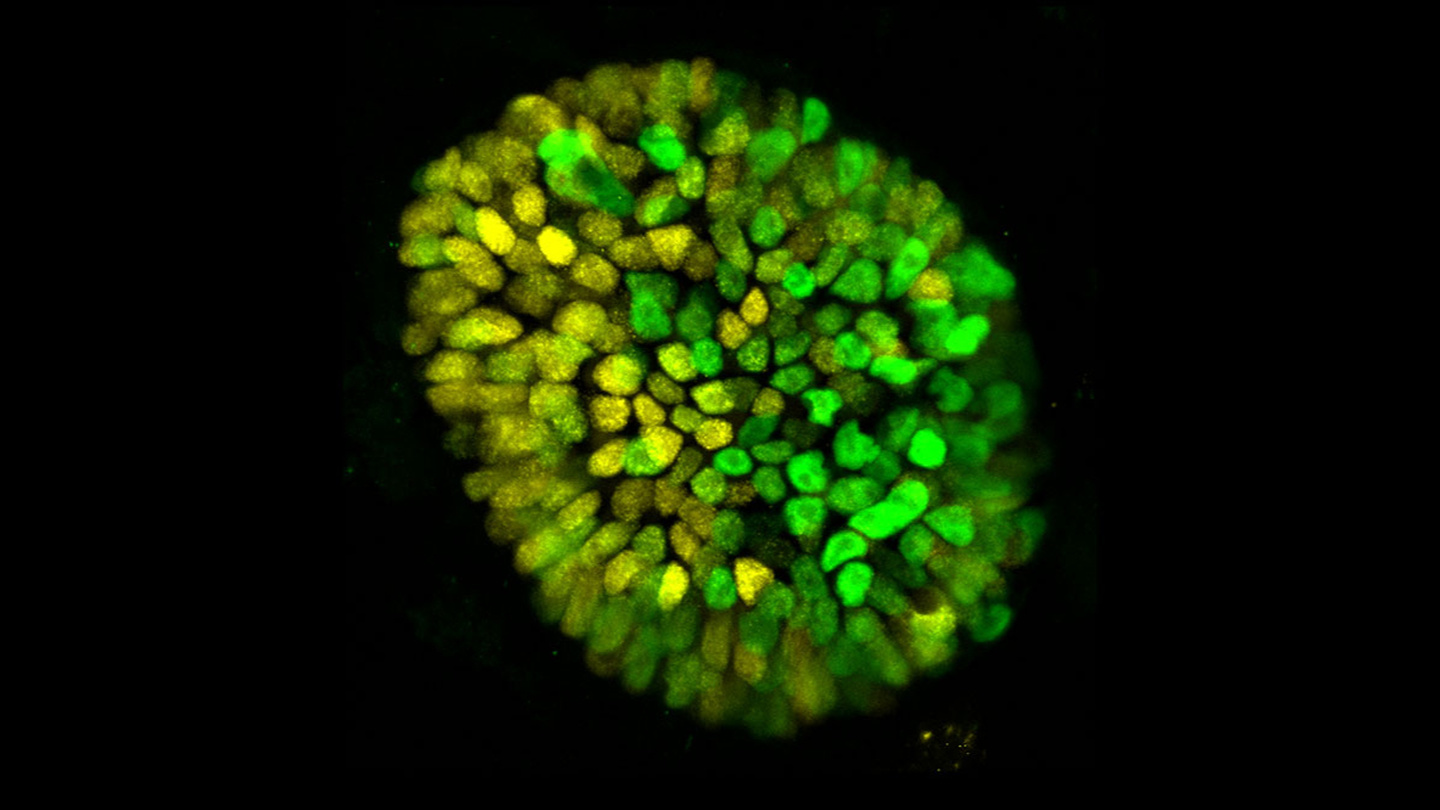
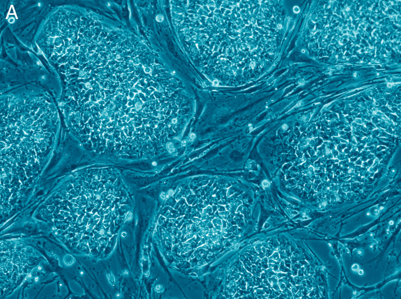
Stems cells have always been pretty amazing.
- New research indicates animals' oldest ancestor was not sponges' single-celled choanocyte bacteria as previously thought.
- It appears our earliest predecessors were something like modern stem cells.
- Our lineage just lost its founding member. The search for our true first predecessor is on!
New technology reveals an old history
<p>The Degnans and their colleagues sequenced the genes expressed by sponge <a href="https://en.wikipedia.org/wiki/Somatic_cell" target="_blank">somatic</a> cell types, which are believed to be similar to those present in the last single-celled common ancestor prior to the emergence of multicellular <a href="https://en.wiktionary.org/wiki/metazoan" target="_blank">metazoans</a> and ultimately, animals. Using the <a href="https://www.ncbi.nlm.nih.gov/pubmed/27121950" target="_blank">CEL-Seq2</a> gene-sequencing platform, they analyzed:</p> <ul> <li>choanocytes — described in the study as "internal <a href="https://en.wikipedia.org/wiki/Epithelium" target="_blank">epithelial</a> feeding cells that capture food by pumping water through the sponge."</li> <li>pinacocytes — epithelial cells that line sponges' internal canals and their outer surface.</li> <li>archaeocytes — <a href="https://en.wikipedia.org/wiki/Mesenchymal_stem_cell" target="_blank">mesenchymal</a> cells from sponges' middle collagenous layer that perform a range of functions.</li> </ul><img type="lazy-image" data-runner-src="https://assets.rebelmouse.io/eyJhbGciOiJIUzI1NiIsInR5cCI6IkpXVCJ9.eyJpbWFnZSI6Imh0dHBzOi8vYXNzZXRzLnJibC5tcy8xOTYwMzYyOS9vcmlnaW4uanBnIiwiZXhwaXJlc19hdCI6MTY3MzEwNTkxOH0.5L1FzU6eitKuQhgOdUrO2sj5TEjfyO0It_tz0I4v4nw/img.jpg?width=980" id="f28e9" class="rm-shortcode" data-rm-shortcode-id="541d4a2fcd2e0adce17994217b068c42" data-rm-shortcode-name="rebelmouse-image" />Image source: QU
Choanocytes are not your great, great, great, etc. grandmother
<p>The researchers found "no support for the long-standing hypothesis that multicellular animals evolved from an ancestor that was an undifferentiated ball of cells resembling extant choanocytes and choanoflagellates." Instead, the truth seemed to lay more in the flexible behavior of archeocytes. The researchers concluded that the "ancestral metazoan cell type had the capacity to exist in and transition between multiple cell states."</p><p>In other words, says Professor Degnan, "We've found that the first multicellular animals probably weren't like the modern-day sponge cells, but were more like a collection of convertible cells. And so, the "great-great-great-grandmother of all cells in the animal kingdom, so to speak, was probably quite similar to a stem cell."</p><p>Associate Professor Degnan notes that modern genetic sequencing tools "helped us finally address an age-old question, discovering something completely contrary to what anyone had ever proposed." As for the exact identity of our earliest single-celled ancestor, she adds, "Now we have an opportunity to re-imagine the steps that gave rise to the first animals, the underlying rules that turned single cells into multicellular animal life."</p><img type="lazy-image" data-runner-src="https://assets.rebelmouse.io/eyJhbGciOiJIUzI1NiIsInR5cCI6IkpXVCJ9.eyJpbWFnZSI6Imh0dHBzOi8vYXNzZXRzLnJibC5tcy8xOTYwMzY1NS9vcmlnaW4uanBnIiwiZXhwaXJlc19hdCI6MTY2MjM4MzkxMX0.Dj0UdR6IFUv0Eq9UWFSIOtUXrVJcEYoHiLSHVzKEQQs/img.jpg?width=980" id="098ac" class="rm-shortcode" data-rm-shortcode-id="91ffa81c64bf7668a9ee976e82cbb0a1" data-rm-shortcode-name="rebelmouse-image" />Amphimedon queenslandica, from which the study's were taken (left), and a choanocyte chamber containing multiple choanocytes (right)
Image sources: Karin Taylor, Rebecca Fieth, QU
The first human esophagus grown in a lab
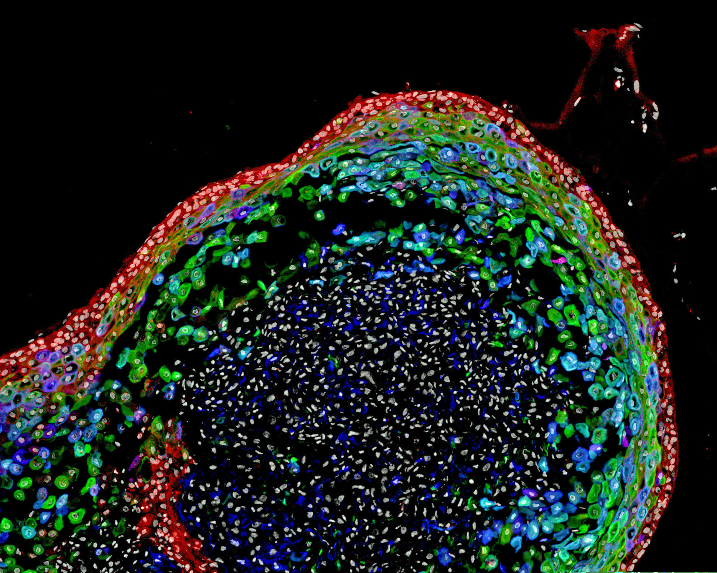
Scientists have grown a model human esophagus using pluripotent stem cells for the first time.
- By precisely timing the application of different chemicals, scientists have grown a small, model esophagus from stem cells.
- They used the model esophagus to clarify why a certain congenital condition occurs.
- Using this technique, future researchers will be able to understand the nature of diseases better, develop new treatments, and even repair damaged esophagi.
Growing an esophagus
<p>Developing this small esophagus organoid took a lot of precision. The 800-micrometer organoid was grown over the period of two months, but it started out as a slurry of pluripotent stem cells (PSCs). Unlike adult stem cells, which can only grow into specific, specialized types of tissues, PSCs can develop into any type of cell in the body. Essentially, they are our original components — every human started off as a similar slurry of PSCs.</p><p>The researchers exposed these cells to precise amounts of different chemicals that recreated the kind of events a PSC would undergo in order to develop into an esophagus in a normal developing fetus. These chemicals manipulated <a href="https://www.cancer.gov/publications/dictionaries/cancer-terms/def/signaling-pathway" target="_blank">cellular signaling pathways</a> — essentially, a chain of reactions that occur when a cell is exposed to a certain molecule. In the cell, a cascading series of reactions occurs that triggers some kind of event in a cell, such as cell death, replication, or, in this case, differentiation into esophagus cells.</p><p>Previous studies had tried to develop human esophagus organoids, but these usually ended up as a mixture of different tissues, including those found in the pharynx, the esophagus, and the respiratory tract. To develop esophageal tissues, the researchers needed to precisely time similarly precise quantities of chemicals to trigger the right signaling pathways for the right amount of time. </p><p>As an example, exposing the cells to retinoic acid for four days caused them to develop into tissues found lower down in the foregut, below the esophagus. Treating the cells in retinoic acid for just one day, however, seemed to be the right amount of exposure to encourage esophageal tissues to develop. In addition, treating the cells with Noggin — a curiously named protein — encouraged the tissues to develop into esophageal tissues rather than respiratory tissues.</p><img type="lazy-image" data-runner-src="https://assets.rebelmouse.io/eyJhbGciOiJIUzI1NiIsInR5cCI6IkpXVCJ9.eyJpbWFnZSI6Imh0dHBzOi8vYXNzZXRzLnJibC5tcy8xODY2OTAyNS9vcmlnaW4uanBnIiwiZXhwaXJlc19hdCI6MTY3MjA4NDk1NH0.fBFBllHrzlJBuy5VqHfk37Fk0oKQcmxPfB4dliINq30/img.jpg?width=980" id="7be54" class="rm-shortcode" data-rm-shortcode-id="5cc12f694599c06332d61fb65b6f0e06" data-rm-shortcode-name="rebelmouse-image" />A diagram depicting the various possible tissues the stem cells could have developed into. Exposing the cells to different molecules, such as retinoic acid (RA) and Noggin (NOG) encourages the stem cells to develop into different tissues.
Trisno et al., 2018
What's useful about this?
<p>Growing a model of the human esophagus is an interesting project, but science like this isn't done out of sheer curiosity. Regarding its utility, Jim Wells, a researcher working on the project, <a href="https://www.sciencedaily.com/releases/2018/09/180920161030.htm" target="_blank">said</a>, "In addition to being a new model to study birth defects like <a href="https://medlineplus.gov/ency/article/000961.htm" target="_blank">esophageal atresia</a>, the organoids can be used to study diseases like <a href="https://medlineplus.gov/eosinophilicesophagitis.html" target="_blank">eosinophilic esophagitis</a> and <a href="https://www.mayoclinic.org/diseases-conditions/barretts-esophagus/symptoms-causes/syc-20352841" target="_blank">Barrett's metaplasia</a>, or to bioengineer genetically matched esophageal tissue for individual patients." There are other potential applications of this research on esophageal cancer, <a href="https://www.medicalnewstoday.com/articles/14085.php" target="_blank">gastroesophageal reflux disease (GERD)</a>, and <a href="https://www.healthline.com/health/achalasia" target="_blank">achalasia</a>, which affects the esophagus's lower muscles, preventing food from passing through. The researchers noted that all of these conditions need better treatments.</p><p>To demonstrate the usefulness of this model organ, the researchers examined the impact of the <em>SOX2</em> gene on the development of the esophagus. In both humans and mice, when <em>SOX2</em> is repressed or inactivated, the esophagus peters out and fails to connect to the stomach. For babies born with esophageal atresia, this condition can be life-threatening and requires surgery to correct.</p><p>Researchers have known that <em>SOX2</em> was associated with this condition, but the exact mechanism was unknown. By studying the growth of the esophageal organoid and comparing it with the esophagi of mice whose <em>Sox2</em> genes had been inactivated, the researchers discovered that a molecule called Wnt was the likely reason behind esophageal atresia. Remember how growing this organoid require precisely timed applications of various chemicals? Wnt works like that to — only in a developing body, the <em>SOX2</em> gene inhibits the amount of Wnt the cells are exposed to. When <em>SOX2</em> doesn't work correctly, Wnt encourages developing cells to become part of the respiratory tract rather than the esophagus, resulting in esophageal atresia.</p><p>This kind of work is very much in the early stages. <a href="https://stemcells.nih.gov/info/basics/1.htm" target="_blank">Before 1998</a>, scientists had no idea how to harvest human stem cells, and now we're building model organs with them. While growing an entire organ is still very much a thing of the future, it's important to remember that every step along the way to that goal will make use better at fighting disease, saving lives, and understanding how the human body functions.</p>Gay couples may soon be able to have their own biological kids
Human egg cells can now be created from donor blood — a brave new world is upon us.
- Japanese scientists have successfully created immature human egg cells using stem cells.
- The discovery builds on years of research into the uses of stem cells.
- While the prospects for new fertility treatments are promising, the ethical questions raised by the procedure will have to be answered.
How did they do it?
<span style="display:block;position:relative;padding-top:56.25%;" class="rm-shortcode" data-rm-shortcode-id="71aae8a9494f6d3b362ec33e10a90089"><iframe type="lazy-iframe" data-runner-src="https://www.youtube.com/embed/DshNfhAwPec?rel=0" width="100%" height="auto" frameborder="0" scrolling="no" style="position:absolute;top:0;left:0;width:100%;height:100%;"></iframe></span><p>The scientists took human blood cells and used a <a href="https://www.hopkinsmedicine.org/news/media/releases/johns_hopkins_researchers_return_blood_cells_to_stem_cell_state" target="_blank">previously known method</a> to transform them into human pluripotent stem cells (hPSCs). These cells can be changed into <a href="https://www.medicalnewstoday.com/info/stem_cell" target="_blank">any other cell,</a> making them extremely useful in medical research and giving them endless potential for <a href="https://bigthink.com/surprising-science/stem-cells-used-to-repair-teeth" target="_blank">future treatments</a>.</p><p>While previous studies have attempted to create lab-grown human eggs from stem cells before, those attempts were all unsuccessful. The trick, it seems, was to put the new egg cells into a miniature ovary built for this task out of cells taken from mice. This allowed the scientists to succeed where others had failed. </p>What can we do with this?
<span style="display:block;position:relative;padding-top:56.25%;" class="rm-shortcode" data-rm-shortcode-id="485e259c7c25c0b4ac0ccb6afcd9986c"><iframe type="lazy-iframe" data-runner-src="https://www.youtube.com/embed/VjY0vqgDMnE?rel=0" width="100%" height="auto" frameborder="0" scrolling="no" style="position:absolute;top:0;left:0;width:100%;height:100%;"></iframe></span><p>While the eggs produced in this study are immature and incapable of doing anything, there is reason to hope that over the long run fully functioning eggs will be <a href="https://www.npr.org/sections/health-shots/2018/09/20/649552734/scientists-create-immature-human-eggs-from-stem-cells" target="_blank">created</a>. This has already been done for <a href="https://www.npr.org/sections/health-shots/2012/10/04/162263750/scientists-create-fertile-eggs-from-mouse-stem-cells" target="_blank">mice</a>, although making that work for human cells will be a little more difficult.</p><p>Artificially created mature human egg cells could be used in a variety of fertility treatments. They could be produced for those born infertile, people who have lost their fertility due to illness, or even for gay couples who want to have a child with their own genetic makeup and not that of a donor. On a more mundane level, mature eggs could be created on demand for research purposes.</p><p>The study's authors stated that the next step for them will be to try and produce mature egg and sperm cells that could actually be used for reproduction. It may only be a matter of time before fully functioning lab grown human gametes are a reality. </p>What are the ethical concerns?
<span style="display:block;position:relative;padding-top:56.25%;" class="rm-shortcode" data-rm-shortcode-id="732941e921e5e20999f80d60e1396aa3"><iframe type="lazy-iframe" data-runner-src="https://www.youtube.com/embed/L558WtD1tU8?rel=0" width="100%" height="auto" frameborder="0" scrolling="no" style="position:absolute;top:0;left:0;width:100%;height:100%;"></iframe></span><p>This process currently allows immature eggs to be created that have the DNA of the blood cell donor. If the procedure is perfected and mature sperm and eggs can be produced, a <a href="https://www.goodreads.com/book/show/5129.Brave_New_World" target="_blank">brave new world</a> of genetic selection may be upon us. One in which desirable DNA can be bought and sold to create gametes with desirable genetic qualities. <a href="https://religion.dartmouth.edu/people/ronald-michael-green" target="_blank">Ronald Green</a>, a Dartmouth bioethicist who often works on issues related to <a href="http://www.washingtonpost.com/wp-dyn/content/article/2008/11/11/AR2008111102258.html" target="_blank">stem cells</a>, explains one of the many strange possibilities that we may soon face:</p><blockquote>A woman might want to have George Clooney's baby, and his hairdresser could start selling his hair follicles online. So we suddenly could see many, many progeny of George Clooney without his consent.</blockquote><p>In addition to the possibility of genetic theft, the ability for any person to have their cells turned into eggs which could lead to a successful birth allows for the creation of children from the cells of the recently deceased. A creepy situation that we will undoubtedly have to reckon with. </p><p>People in need of new organs might see fit to have a child created with their DNA to provide a spare kidney a few years down the road. This is already happening in the form of ethically ambiguous <a href="http://www.amc.edu/bioethicsblog/post.cfm/is-it-ethical-for-parents-to-create-a-savior-sibling" target="_blank">savior siblings</a> and was explored in the book and film <a href="https://www.imdb.com/title/tt1078588/" target="_blank"><em>My Sister's Keeper</em></a><em>. </em>Will the morality of the child's creation be any different if the child is tailor made for the person in need of a donor rather than the old fashioned way? </p><p>We must begin asking ourselves now how we will react to these ethical questions since the science won't slow down for us.</p>Human skeletal stem cells isolated in breakthrough discovery
It's a development that could one day lead to much better treatments for osteoporosis, joint damage, and bone fractures.
- Scientists have isolated skeletal stem cells in adult and fetal bones for the first time.
- These cells could one day help treat damaged bone and cartilage.
- The team was able to grow skeletal stem cells from cells found within liposuctioned fat.
Isolating skeletal stem cells in humans
<p>Still, it wasn't clear these skeletal stem cells existed in humans because we're vastly more complex than mice. To find out, the team searched fetal bones for stem cells with a genetic signature similar to that of the skeletal stem cells in mice. After isolating these stem cells in lab dishes, they reliably grew into only bone, cartilage, and stroma (essentially, a mix of connective tissue and blood vessels).<br></p><p>The researchers confirmed that these cells were indeed dedicated human skeletal stem cells by isolating the same type of cells from adult human bones, obtained from fragments that had been recently removed during hip surgeries. Again, these stem cells only produced bone, cartilage, and stroma. </p><p>What's more, the researchers were able to produce human skeletal stem cells from stromal cells. As <a href="https://www.sciencemag.org/news/2018/09/skeletal-stem-cells-found-humans-first-time-promising-new-treatments-fractures-and" target="_blank"><em>Science</em></a> reports, they accomplished this by isolating stromal cells and growing them alongside a bone-growth protein. </p><p>It turns out there's an abundant potential source for stromal cells: liposuctioned fat. </p><p>"A half-million times a year, U.S. citizens have their fat sucked out and it's discarded as medical waste," study author Michael Longaker of Stanford University told <em>Science</em>. "That's a lot of material we could use to generate skeletal stem cells." </p><p>Although it could take years before these cells are used to treat joint damage or replace broken bones, the discovery provides a proof-of-concept for future treatments of degenerative skeletal conditions like osteoporosis, which <a href="https://www.iofbonehealth.org/facts-statistics" target="_blank">affects 44 million Americans.</a></p>