Big ideas.
Once a week.
Subscribe to our weekly newsletter.
Where does intelligence reside in the brain?
There are a few different theories out there, but the parieto-frontal integration theory, or P-FIT, appears to give us the best model of the neuroscience of intelligence.

- Intelligence is such a complex phenomenon that it's difficult to imagine specific regions of the brain could be responsible for greater, lesser, or different varieties of intelligence.
- However, neuroimaging and brain lesion research has enabled us to pinpoint the neural networks that are most likely to be involved in intelligence.
- The best contender thus far is the parieto-frontal integration theory, or P-FIT, although other models of the neuroscience of intelligence exist.
For some, the concept that our brains — the simple, three-pound chunks of matter we carry around in our skulls — could determine who we are is a distressing one. The idea of having an ethereal, eternal soul is a much more comforting thought, and somehow, it feels more reasonable to assert than that a complex experience as consciousness could arise from something as mundane as an organ.
But when you zoom in far enough, this proposal becomes less fantastical: the brain contains about 86 billion neurons, with over 100 trillion connections between those neurons. When you consider this density of connections, it's clear that some pretty impressive phenomenon can come out of a measly three pounds. Although consciousness is a multifaceted experience, neuroscientists are increasingly able to pin down which regions of the brain correspond to which phenomenon in our daily life. For instance, neuroscientists have a pretty good idea of where intelligence — arguably one of the most fundamental features of consciousness — resides in our brains.
Tracking down intelligence in the brain
In the early 20th century, a psychologist named Charles Spearman discovered an interesting and intuitive correlation. Students who performed well in one subject tended to perform well in other, completely unrelated subjects. There was never a negative correlation between performance; a student gifted in reading also tended to perform well at math, though maybe less spectacularly. This suggested a hidden factor driving success in all these disparate domains.
Spearman called this factor g — or, the general intelligence factor. It's what we think of when we think of what intelligence is, a kind of all-around capacity for understanding and integrating information and using it to solve problems. An individual who is particularly gifted at mathematics will likely perform well in memory retrieval tasks, in recognizing patterns, in reading comprehension, and so on because they have a high g factor. IQ scores are primarily derived from this specific factor, rather than merely one cognitive domain or the other.
For a long time, neuroscientists struggled with the puzzle of which part of our brains gave rise to general intelligence. We knew that the sensation of fear arose in the amygdala, that the hippocampus has a great deal to do with the encoding of memories, and even that the prefrontal cortex is associated with our executive functioning, such as our ability to switch between tasks and inhibit our behaviors. However, although the prefrontal cortex certainly has a lot to do with intelligence, the specific area or areas of the brain that determine how well somebody can recognize patterns, think critically about a subject, or speak persuasively were a mystery for a long time.
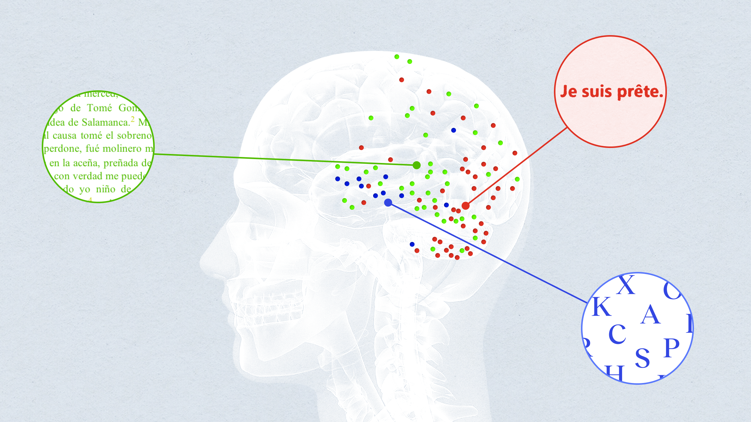
Now, thanks to neuroimaging research, many neuroscientists believe that a network across several brain regions is responsible for intelligence. The parieto-frontal integration theory, or P-FIT, proposes that intelligence arises from a network mostly located in the frontal and parietal lobes.
Image depicting regions of the parieto-frontal network. The numbers refer to specific Brodmann's areas, or regions of the brain defined by their cellular structure. Dark-colored circles are primarily associated with the left hemisphere, while light-colored circles are primarily associated with the left hemisphere. The white arrow refers to the arcuate fasciculus, a bundle of nerve fibers connecting parts of the brain, notably Wernicke's and Broca's areas.
Jung & Haier, 2007.
In a review of 37 neuroimaging studies focused on intelligence and conducted on more than 1,500 individuals, researchers found that activity in the parieto-frontal network correlated with higher scores on the Wechsler Adult Intelligence Scale for the majority of participants. The researchers identified that the network's efficiency in communication was responsible for an individual being more or less intelligent, and variations in this network also corresponded to the differences in how individuals approach problems.
"Recent neuroscience studies suggest that intelligence is related to how well information travels throughout the brain," said Richard Haier in a press release, a professor of psychology at the University of California, Irvine, who helped identify the parieto-frontal network. "Our review of imaging studies identifies the stations along the routes intelligent information processing takes. Once we know where the stations are, we can study how they relate to intelligence."
How well does P-FIT fit?
However, this theory is mainly based on images from fMRIs, which are problematic in a few ways. The first is that they are correlational. It's difficult to definitively claim that because one region of the brain lights up under an fMRI when a task is being performed, that region is responsible for task performance.
In addition, modern fMRIs can track blood flow to different regions of the brain, but they don't actually track the activity of specific neurons. Neuron activity and blood flow are linked, but since we can only measure blood flow, we lose some details that may provide insight.
That's why studying brain lesions are an important part of neuroscience. Lesions are just any kind of tissue damage, and brain lesions are useful to researchers because they can show definitively what functions that region of the brain has. For instance, when a lesion occurs in Wernicke's region in the brain, the affected individual will have difficulty comprehending language but will remain capable of speech; this is because Wernicke's region deals with language comprehension, while Broca's region deals with the production of language. When researchers have had the opportunity to study brain lesions in the regions identified by P-FIT, they seem to confirm the theory.
For example, researchers scanned the brains of 182 Vietnam War veterans who had received brain damage during the war. They identified the locations of their brain lesions and administered the Weschler Adult Intelligence Scale and the Delis-Kaplan Executive Function System, which measure IQ and executive function, respectively. The results found that IQ and executive function scores did, indeed, vary depending on whether participants' lesions were located in the parieto-frontal network or not.
The case is still open
Of course, P-FIT is far from the only theory we have about intelligence. Some, for instance, argue that brain waves are the most influential component of intelligence, coordinating neural activity depending on the task at hand. Others claim that rather than any one network in the brain, intelligence is more a function of neural plasticity and the dynamic reorganization of brain networks — the more plasticky and flexible your brain is, the smarter you are.
We're still a long way off from knowing for certain which parts of the brain create intelligence, or whether this is even a reasonable puzzle to solve. Thus far, P-FIT seems to be the strongest theory around, although its competitors have similarly compelling cases. As our technology and understanding develops, hopefully we'll have a more complete picture of intelligence in the future.
Weird science shows unseemly way beetles escape after being eaten
Certain water beetles can escape from frogs after being consumed.
R. attenuata escaping from a black-spotted pond frog.
- A Japanese scientist shows that some beetles can wiggle out of frog's butts after being eaten whole.
- The research suggests the beetle can get out in as little as 7 minutes.
- Most of the beetles swallowed in the experiment survived with no complications after being excreted.
In what is perhaps one of the weirdest experiments ever that comes from the category of "why did anyone need to know this?" scientists have proven that the Regimbartia attenuata beetle can climb out of a frog's butt after being eaten.
The research was carried out by Kobe University ecologist Shinji Sugiura. His team found that the majority of beetles swallowed by black-spotted pond frogs (Pelophylax nigromaculatus) used in their experiment managed to escape about 6 hours after and were perfectly fine.
"Here, I report active escape of the aquatic beetle R. attenuata from the vents of five frog species via the digestive tract," writes Sugiura in a new paper, adding "although adult beetles were easily eaten by frogs, 90 percent of swallowed beetles were excreted within six hours after being eaten and, surprisingly, were still alive."
One bug even got out in as little as 7 minutes.
Sugiura also tried putting wax on the legs of some of the beetles, preventing them from moving. These ones were not able to make it out alive, taking from 38 to 150 hours to be digested.
Naturally, as anyone would upon encountering such a story, you're wondering where's the video. Thankfully, the scientists recorded the proceedings:
The Regimbartia attenuata beetle can be found in the tropics, especially as pests in fish hatcheries. It's not the only kind of creature that can survive being swallowed. A recent study showed that snake eels are able to burrow out of the stomachs of fish using their sharp tails, only to become stuck, die, and be mummified in the gut cavity. Scientists are calling the beetle's ability the first documented "active prey escape." Usually, such travelers through the digestive tract have particular adaptations that make it possible for them to withstand extreme pH and lack of oxygen. The researchers think the beetle's trick is in inducing the frog to open a so-called "vent" controlled by the sphincter muscle.
"Individuals were always excreted head first from the frog vent, suggesting that R. attenuata stimulates the hind gut, urging the frog to defecate," explains Sugiura.
For more information, check out the study published in Current Biology.
We're creating pigs with human immune systems to study illness
Are "humanized" pigs the future of medical research?
The U.S. Food and Drug Administration requires all new medicines to be tested in animals before use in people. Pigs make better medical research subjects than mice, because they are closer to humans in size, physiology and genetic makeup.
In recent years, our team at Iowa State University has found a way to make pigs an even closer stand-in for humans. We have successfully transferred components of the human immune system into pigs that lack a functional immune system. This breakthrough has the potential to accelerate medical research in many areas, including virus and vaccine research, as well as cancer and stem cell therapeutics.
Existing biomedical models
Severe Combined Immunodeficiency, or SCID, is a genetic condition that causes impaired development of the immune system. People can develop SCID, as dramatized in the 1976 movie “The Boy in the Plastic Bubble." Other animals can develop SCID, too, including mice.
Researchers in the 1980s recognized that SCID mice could be implanted with human immune cells for further study. Such mice are called “humanized" mice and have been optimized over the past 30 years to study many questions relevant to human health.
Mice are the most commonly used animal in biomedical research, but results from mice often do not translate well to human responses, thanks to differences in metabolism, size and divergent cell functions compared with people.
Nonhuman primates are also used for medical research and are certainly closer stand-ins for humans. But using them for this purpose raises numerous ethical considerations. With these concerns in mind, the National Institutes of Health retired most of its chimpanzees from biomedical research in 2013.
Alternative animal models are in demand.
Swine are a viable option for medical research because of their similarities to humans. And with their widespread commercial use, pigs are met with fewer ethical dilemmas than primates. Upwards of 100 million hogs are slaughtered each year for food in the U.S.
Humanizing pigs
In 2012, groups at Iowa State University and Kansas State University, including Jack Dekkers, an expert in animal breeding and genetics, and Raymond Rowland, a specialist in animal diseases, serendipitously discovered a naturally occurring genetic mutation in pigs that caused SCID. We wondered if we could develop these pigs to create a new biomedical model.
Our group has worked for nearly a decade developing and optimizing SCID pigs for applications in biomedical research. In 2018, we achieved a twofold milestone when working with animal physiologist Jason Ross and his lab. Together we developed a more immunocompromised pig than the original SCID pig – and successfully humanized it, by transferring cultured human immune stem cells into the livers of developing piglets.
During early fetal development, immune cells develop within the liver, providing an opportunity to introduce human cells. We inject human immune stem cells into fetal pig livers using ultrasound imaging as a guide. As the pig fetus develops, the injected human immune stem cells begin to differentiate – or change into other kinds of cells – and spread through the pig's body. Once SCID piglets are born, we can detect human immune cells in their blood, liver, spleen and thymus gland. This humanization is what makes them so valuable for testing new medical treatments.
We have found that human ovarian tumors survive and grow in SCID pigs, giving us an opportunity to study ovarian cancer in a new way. Similarly, because human skin survives on SCID pigs, scientists may be able to develop new treatments for skin burns. Other research possibilities are numerous.
The ultraclean SCID pig biocontainment facility in Ames, Iowa. Adeline Boettcher, CC BY-SA
Pigs in a bubble
Since our pigs lack essential components of their immune system, they are extremely susceptible to infection and require special housing to help reduce exposure to pathogens.
SCID pigs are raised in bubble biocontainment facilities. Positive pressure rooms, which maintain a higher air pressure than the surrounding environment to keep pathogens out, are coupled with highly filtered air and water. All personnel are required to wear full personal protective equipment. We typically have anywhere from two to 15 SCID pigs and breeding animals at a given time. (Our breeding animals do not have SCID, but they are genetic carriers of the mutation, so their offspring may have SCID.)
As with any animal research, ethical considerations are always front and center. All our protocols are approved by Iowa State University's Institutional Animal Care and Use Committee and are in accordance with The National Institutes of Health's Guide for the Care and Use of Laboratory Animals.
Every day, twice a day, our pigs are checked by expert caretakers who monitor their health status and provide engagement. We have veterinarians on call. If any pigs fall ill, and drug or antibiotic intervention does not improve their condition, the animals are humanely euthanized.
Our goal is to continue optimizing our humanized SCID pigs so they can be more readily available for stem cell therapy testing, as well as research in other areas, including cancer. We hope the development of the SCID pig model will pave the way for advancements in therapeutic testing, with the long-term goal of improving human patient outcomes.
Adeline Boettcher earned her research-based Ph.D. working on the SCID project in 2019.
Christopher Tuggle, Professor of Animal Science, Iowa State University and Adeline Boettcher, Technical Writer II, Iowa State University
This article is republished from The Conversation under a Creative Commons license. Read the original article.
A new warning to sign to predict volcanic eruptions?
Satellite imagery can help better predict volcanic eruptions by monitoring changes in surface temperature near volcanoes.
Volcano erupting lava, volcanic sky active rock night Ecuador landscape
- A recent study used data collected by NASA satellites to conduct a statistical analysis of surface temperatures near volcanoes that erupted from 2002 to 2019.
- The results showed that surface temperatures near volcanoes gradually increased in the months and years prior to eruptions.
- The method was able to detect potential eruptions that were not anticipated by other volcano monitoring methods, such as eruptions in Japan in 2014 and Chile in 2015.
How can modern technology help warn us of impending volcanic eruptions?
One promising answer may lie in satellite imagery. In a recent study published in Nature Geoscience, researchers used infrared data collected by NASA satellites to study the conditions near volcanoes in the months and years before they erupted.
The results revealed a pattern: Prior to eruptions, an unusually large amount of heat had been escaping through soil near volcanoes. This diffusion of subterranean heat — which is a byproduct of "large-scale thermal unrest" — could potentially represent a warning sign of future eruptions.
Conceptual model of large-scale thermal unrestCredit: Girona et al.
For the study, the researchers conducted a statistical analysis of changes in surface temperature near volcanoes, using data collected over 16.5 years by NASA's Terra and Aqua satellites. The results showed that eruptions tended to occur around the time when surface temperatures near the volcanoes peaked.
Eruptions were preceded by "subtle but significant long-term (years), large-scale (tens of square kilometres) increases in their radiant heat flux (up to ~1 °C in median radiant temperature)," the researchers wrote. After eruptions, surface temperatures reliably decreased, though the cool-down period took longer for bigger eruptions.
"Volcanoes can experience thermal unrest for several years before eruption," the researchers wrote. "This thermal unrest is dominated by a large-scale phenomenon operating over extensive areas of volcanic edifices, can be an early indicator of volcanic reactivation, can increase prior to different types of eruption and can be tracked through a statistical analysis of little-processed (that is, radiance or radiant temperature) satellite-based remote sensing data with high temporal resolution."
Temporal variations of target volcanoesCredit: Girona et al.
Although using satellites to monitor thermal unrest wouldn't enable scientists to make hyper-specific eruption predictions (like predicting the exact day), it could significantly improve prediction efforts. Seismologists and volcanologists currently use a range of techniques to forecast eruptions, including monitoring for gas emissions, ground deformation, and changes to nearby water channels, to name a few.
Still, none of these techniques have proven completely reliable, both because of the science and the practical barriers (e.g. funding) standing in the way of large-scale monitoring. In 2014, for example, Japan's Mount Ontake suddenly erupted, killing 63 people. It was the nation's deadliest eruption in nearly a century.
In the study, the researchers found that surface temperatures near Mount Ontake had been increasing in the two years prior to the eruption. To date, no other monitoring method has detected "well-defined" warning signs for the 2014 disaster, the researchers noted.
The researchers hope satellite-based infrared monitoring techniques, combined with existing methods, can improve prediction efforts for volcanic eruptions. Volcanic eruptions have killed about 2,000 people since 2000.
"Our findings can open new horizons to better constrain magma–hydrothermal interaction processes, especially when integrated with other datasets, allowing us to explore the thermal budget of volcanoes and anticipate eruptions that are very difficult to forecast through other geophysical/geochemical methods."
Moral and economic lessons from Mario Kart
The design of a classic video game yields insights on how to address global poverty.