Big ideas.
Once a week.
Subscribe to our weekly newsletter.
This Is Your Brain During Orgasm
If you ever want to make even the most cosmopolitan of your friends speechless, telling them you have volunteered to travel to Newark, New Jersey, so you can masturbate to orgasm in an fMRI is a great way to start. Once they overcome the shock, chances are they will start to ask questions. Most I was able to answer.

Editor's Note: A writer by trade, Kayt Sukel volunteered to masturbate in an MRI scanner for science. The point of the study? Neuroscientist Barry Komisaruk and sex therapist Nan Wise wanted to know what exactly goes on in the brain when a woman orgasms. Could the sensory cortex be activated by thought alone, they wondered, opening doors for treatments for people unable to orgasm through genital stimulation? This guest post is an excerpt from Sukel's just-released Dirty Minds: How Our Brains Influence Love, Sex, and Relationships.
What's the Big Idea?
If you ever want to make even the most cosmopolitan of your friends speechless, telling them you have volunteered to travel to Newark, New Jersey, so you can masturbate to orgasm in an fMRI is a great way to start.
Once they overcome the shock, chances are they will start to ask questions. Most I was able to answer. To start, no, I’m not kidding, I’m really going to do it. Yes, I will be in the scanner, the same sort of claustrophobic tube you got your knee scanned in that one time. Yes, I know it is a very tight fit. Loud too. Yes, I’ll be self-stimulating. How? Clitorally, to be exact, until I reach orgasm. Will I use a vibrator? No, most vibrators have metal, which is a no-no in the magnet.
I was going through the same spiel over and over again. I knew the procedure backward and forward. Or so I thought. When I arrived at Rutgers University’s Smith Hall, a 1970s-style building in the middle of the Newark campus, I was in a bit of a panic. Despite spending an hour or two trying to concoct some kind of sexy fantasy about lab coats and confined spaces the previous night, I was afraid that when push came to shove, I would not be able to reach orgasm.
The first order of business was to fit me for a head mask, a sort of modern Count of Monte Cristo–type restraint system made of tight plastic mesh. White and blue, the contraption was part low-budget bondage porn prop and part clinical radiation treatment kit. Once we started the scan, it would be screwed directly to the scanner bed, meaning that I would be unable to get into or out of the fMRI tube without assistance. Ah. No pressure then. None at all.
Getting to the Big O
A few hours later the party moved to the fMRI suite at the nearby University of Medicine and Dentistry of New Jersey Medical Center. I donned a hospital johnny and was pushed back into the scanner’s tube, as ready as I would ever be to have an orgasm in an fMRI. The magnet started to spin around me. As promised, it was loud. It lasted the majority of my session inside the scanner, which was approximately an hour and a half. Even with ear protection, I could feel each click, clank, and whir all the way down my spine.
Just as I was starting to zone out, not into sleep exactly, but into something like it, the noises suddenly stopped. It was now time for the big show. Ready or not, I had to woman up and bring myself to orgasm. In a few minutes I would know if loud clanks and clicks, hospital johnnies, and a tight mesh head restraint could make the magic happen.
Hearing my cue, I took a deep breath and got to it. It may not have been romantic or sexy in there and, man, this mask thing was starting to get really uncomfortable, but I was going to orgasm no matter what. I powered through it, keeping my head as still as possible. A few minutes later I raised my hand to let Komisaruk know my orgasm had begun. I wouldn’t say it was one of my best, but, hey, in my humble opinion, it still qualified.
I lowered my hand to signal my finish and, with it, let out a long breath of relief. If I could have reached around to pat myself on the back, I would have done so. I now had a great story if anyone ever asked me to name the strangest place I’ve ever had an orgasm. And I had helped science while doing it. Triumph for all parties concerned.
“An Orgasm Is a Whole Brain Experience.”
Two months later, in sunny San Diego, I met Komisaruk and Wise at the Society for Neuroscience conference, which gathers approximately 30,000 neuroscientists to discuss the newest advances in the field. They presented the data from my time in the magnet. And they did so with a 3-D movie highlighting the time line of brain activation. (Call it brain porn.) As I watched the film I was struck by the sheer amount of activation.
What happened in my brain during orgasm? Komisaruk and his colleagues saw distinct temporal activity, with different brain areas being recruited as I went from arousal to orgasm and then back around again to rest.
As I roughed up the suspect, so to speak, my genital sensory cortex, motor areas, hypothalamus, thalamus, and substantia nigra lit up. The hypothalamus was no surprise; it has consistently been implicated in all manner of reproductive behaviors, including arousal. The part of the brain that produces oxytocin, is located there too. My motor areas controlled my fingers as I self-stimulated, and my genital sensory cortex registered that stimulation. And the thalamus? It was integrating not only the activity of my wandering fingers but also the memories and fantasies I used to help build up my arousal. The substantia nigra, an area rich in dopamine-producing neurons, paired with the PVN’s oxytocin release, had me feeling nice and relaxed.
Areas implicated in memory, integration of sensory information, and emotion also became active. As my orgasm came to a close, the hypothalamus turned back on, and reward areas like the nucleus accumbens and caudate nucleus were flooded with dopamine. That was what gave me that final rush. Getting to that point involves a variety of cognitive, emotional, and sensory components—even when it’s just you doing the work.
What's the Significance?
Like every study, Komisaruk's research raises more questions than it answers. Researchers still hope to understand:
As Komisaruk told Sukel: “I can envision a time when people can regulate their own brain chemistry through some kind of internal process. But we’re still in the infancy. Hell, we’re still in the prenatal in this field. But I can’t wait to see what will come in the next ten years. It’s going to be amazing.”
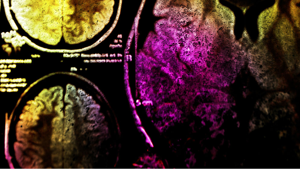
Image courtesy of Shutterstock.
Excerpt courtesy of Free Press. To read more, check out Sukel's book.
Weird science shows unseemly way beetles escape after being eaten
Certain water beetles can escape from frogs after being consumed.
R. attenuata escaping from a black-spotted pond frog.
- A Japanese scientist shows that some beetles can wiggle out of frog's butts after being eaten whole.
- The research suggests the beetle can get out in as little as 7 minutes.
- Most of the beetles swallowed in the experiment survived with no complications after being excreted.
In what is perhaps one of the weirdest experiments ever that comes from the category of "why did anyone need to know this?" scientists have proven that the Regimbartia attenuata beetle can climb out of a frog's butt after being eaten.
The research was carried out by Kobe University ecologist Shinji Sugiura. His team found that the majority of beetles swallowed by black-spotted pond frogs (Pelophylax nigromaculatus) used in their experiment managed to escape about 6 hours after and were perfectly fine.
"Here, I report active escape of the aquatic beetle R. attenuata from the vents of five frog species via the digestive tract," writes Sugiura in a new paper, adding "although adult beetles were easily eaten by frogs, 90 percent of swallowed beetles were excreted within six hours after being eaten and, surprisingly, were still alive."
One bug even got out in as little as 7 minutes.
Sugiura also tried putting wax on the legs of some of the beetles, preventing them from moving. These ones were not able to make it out alive, taking from 38 to 150 hours to be digested.
Naturally, as anyone would upon encountering such a story, you're wondering where's the video. Thankfully, the scientists recorded the proceedings:
The Regimbartia attenuata beetle can be found in the tropics, especially as pests in fish hatcheries. It's not the only kind of creature that can survive being swallowed. A recent study showed that snake eels are able to burrow out of the stomachs of fish using their sharp tails, only to become stuck, die, and be mummified in the gut cavity. Scientists are calling the beetle's ability the first documented "active prey escape." Usually, such travelers through the digestive tract have particular adaptations that make it possible for them to withstand extreme pH and lack of oxygen. The researchers think the beetle's trick is in inducing the frog to open a so-called "vent" controlled by the sphincter muscle.
"Individuals were always excreted head first from the frog vent, suggesting that R. attenuata stimulates the hind gut, urging the frog to defecate," explains Sugiura.
For more information, check out the study published in Current Biology.
We're creating pigs with human immune systems to study illness
Are "humanized" pigs the future of medical research?
The U.S. Food and Drug Administration requires all new medicines to be tested in animals before use in people. Pigs make better medical research subjects than mice, because they are closer to humans in size, physiology and genetic makeup.
In recent years, our team at Iowa State University has found a way to make pigs an even closer stand-in for humans. We have successfully transferred components of the human immune system into pigs that lack a functional immune system. This breakthrough has the potential to accelerate medical research in many areas, including virus and vaccine research, as well as cancer and stem cell therapeutics.
Existing biomedical models
Severe Combined Immunodeficiency, or SCID, is a genetic condition that causes impaired development of the immune system. People can develop SCID, as dramatized in the 1976 movie “The Boy in the Plastic Bubble." Other animals can develop SCID, too, including mice.
Researchers in the 1980s recognized that SCID mice could be implanted with human immune cells for further study. Such mice are called “humanized" mice and have been optimized over the past 30 years to study many questions relevant to human health.
Mice are the most commonly used animal in biomedical research, but results from mice often do not translate well to human responses, thanks to differences in metabolism, size and divergent cell functions compared with people.
Nonhuman primates are also used for medical research and are certainly closer stand-ins for humans. But using them for this purpose raises numerous ethical considerations. With these concerns in mind, the National Institutes of Health retired most of its chimpanzees from biomedical research in 2013.
Alternative animal models are in demand.
Swine are a viable option for medical research because of their similarities to humans. And with their widespread commercial use, pigs are met with fewer ethical dilemmas than primates. Upwards of 100 million hogs are slaughtered each year for food in the U.S.
Humanizing pigs
In 2012, groups at Iowa State University and Kansas State University, including Jack Dekkers, an expert in animal breeding and genetics, and Raymond Rowland, a specialist in animal diseases, serendipitously discovered a naturally occurring genetic mutation in pigs that caused SCID. We wondered if we could develop these pigs to create a new biomedical model.
Our group has worked for nearly a decade developing and optimizing SCID pigs for applications in biomedical research. In 2018, we achieved a twofold milestone when working with animal physiologist Jason Ross and his lab. Together we developed a more immunocompromised pig than the original SCID pig – and successfully humanized it, by transferring cultured human immune stem cells into the livers of developing piglets.
During early fetal development, immune cells develop within the liver, providing an opportunity to introduce human cells. We inject human immune stem cells into fetal pig livers using ultrasound imaging as a guide. As the pig fetus develops, the injected human immune stem cells begin to differentiate – or change into other kinds of cells – and spread through the pig's body. Once SCID piglets are born, we can detect human immune cells in their blood, liver, spleen and thymus gland. This humanization is what makes them so valuable for testing new medical treatments.
We have found that human ovarian tumors survive and grow in SCID pigs, giving us an opportunity to study ovarian cancer in a new way. Similarly, because human skin survives on SCID pigs, scientists may be able to develop new treatments for skin burns. Other research possibilities are numerous.
The ultraclean SCID pig biocontainment facility in Ames, Iowa. Adeline Boettcher, CC BY-SA
Pigs in a bubble
Since our pigs lack essential components of their immune system, they are extremely susceptible to infection and require special housing to help reduce exposure to pathogens.
SCID pigs are raised in bubble biocontainment facilities. Positive pressure rooms, which maintain a higher air pressure than the surrounding environment to keep pathogens out, are coupled with highly filtered air and water. All personnel are required to wear full personal protective equipment. We typically have anywhere from two to 15 SCID pigs and breeding animals at a given time. (Our breeding animals do not have SCID, but they are genetic carriers of the mutation, so their offspring may have SCID.)
As with any animal research, ethical considerations are always front and center. All our protocols are approved by Iowa State University's Institutional Animal Care and Use Committee and are in accordance with The National Institutes of Health's Guide for the Care and Use of Laboratory Animals.
Every day, twice a day, our pigs are checked by expert caretakers who monitor their health status and provide engagement. We have veterinarians on call. If any pigs fall ill, and drug or antibiotic intervention does not improve their condition, the animals are humanely euthanized.
Our goal is to continue optimizing our humanized SCID pigs so they can be more readily available for stem cell therapy testing, as well as research in other areas, including cancer. We hope the development of the SCID pig model will pave the way for advancements in therapeutic testing, with the long-term goal of improving human patient outcomes.
Adeline Boettcher earned her research-based Ph.D. working on the SCID project in 2019.
Christopher Tuggle, Professor of Animal Science, Iowa State University and Adeline Boettcher, Technical Writer II, Iowa State University
This article is republished from The Conversation under a Creative Commons license. Read the original article.
A new warning to sign to predict volcanic eruptions?
Satellite imagery can help better predict volcanic eruptions by monitoring changes in surface temperature near volcanoes.
Volcano erupting lava, volcanic sky active rock night Ecuador landscape
- A recent study used data collected by NASA satellites to conduct a statistical analysis of surface temperatures near volcanoes that erupted from 2002 to 2019.
- The results showed that surface temperatures near volcanoes gradually increased in the months and years prior to eruptions.
- The method was able to detect potential eruptions that were not anticipated by other volcano monitoring methods, such as eruptions in Japan in 2014 and Chile in 2015.
How can modern technology help warn us of impending volcanic eruptions?
One promising answer may lie in satellite imagery. In a recent study published in Nature Geoscience, researchers used infrared data collected by NASA satellites to study the conditions near volcanoes in the months and years before they erupted.
The results revealed a pattern: Prior to eruptions, an unusually large amount of heat had been escaping through soil near volcanoes. This diffusion of subterranean heat — which is a byproduct of "large-scale thermal unrest" — could potentially represent a warning sign of future eruptions.
Conceptual model of large-scale thermal unrestCredit: Girona et al.
For the study, the researchers conducted a statistical analysis of changes in surface temperature near volcanoes, using data collected over 16.5 years by NASA's Terra and Aqua satellites. The results showed that eruptions tended to occur around the time when surface temperatures near the volcanoes peaked.
Eruptions were preceded by "subtle but significant long-term (years), large-scale (tens of square kilometres) increases in their radiant heat flux (up to ~1 °C in median radiant temperature)," the researchers wrote. After eruptions, surface temperatures reliably decreased, though the cool-down period took longer for bigger eruptions.
"Volcanoes can experience thermal unrest for several years before eruption," the researchers wrote. "This thermal unrest is dominated by a large-scale phenomenon operating over extensive areas of volcanic edifices, can be an early indicator of volcanic reactivation, can increase prior to different types of eruption and can be tracked through a statistical analysis of little-processed (that is, radiance or radiant temperature) satellite-based remote sensing data with high temporal resolution."
Temporal variations of target volcanoesCredit: Girona et al.
Although using satellites to monitor thermal unrest wouldn't enable scientists to make hyper-specific eruption predictions (like predicting the exact day), it could significantly improve prediction efforts. Seismologists and volcanologists currently use a range of techniques to forecast eruptions, including monitoring for gas emissions, ground deformation, and changes to nearby water channels, to name a few.
Still, none of these techniques have proven completely reliable, both because of the science and the practical barriers (e.g. funding) standing in the way of large-scale monitoring. In 2014, for example, Japan's Mount Ontake suddenly erupted, killing 63 people. It was the nation's deadliest eruption in nearly a century.
In the study, the researchers found that surface temperatures near Mount Ontake had been increasing in the two years prior to the eruption. To date, no other monitoring method has detected "well-defined" warning signs for the 2014 disaster, the researchers noted.
The researchers hope satellite-based infrared monitoring techniques, combined with existing methods, can improve prediction efforts for volcanic eruptions. Volcanic eruptions have killed about 2,000 people since 2000.
"Our findings can open new horizons to better constrain magma–hydrothermal interaction processes, especially when integrated with other datasets, allowing us to explore the thermal budget of volcanoes and anticipate eruptions that are very difficult to forecast through other geophysical/geochemical methods."
Moral and economic lessons from Mario Kart
The design of a classic video game yields insights on how to address global poverty.
