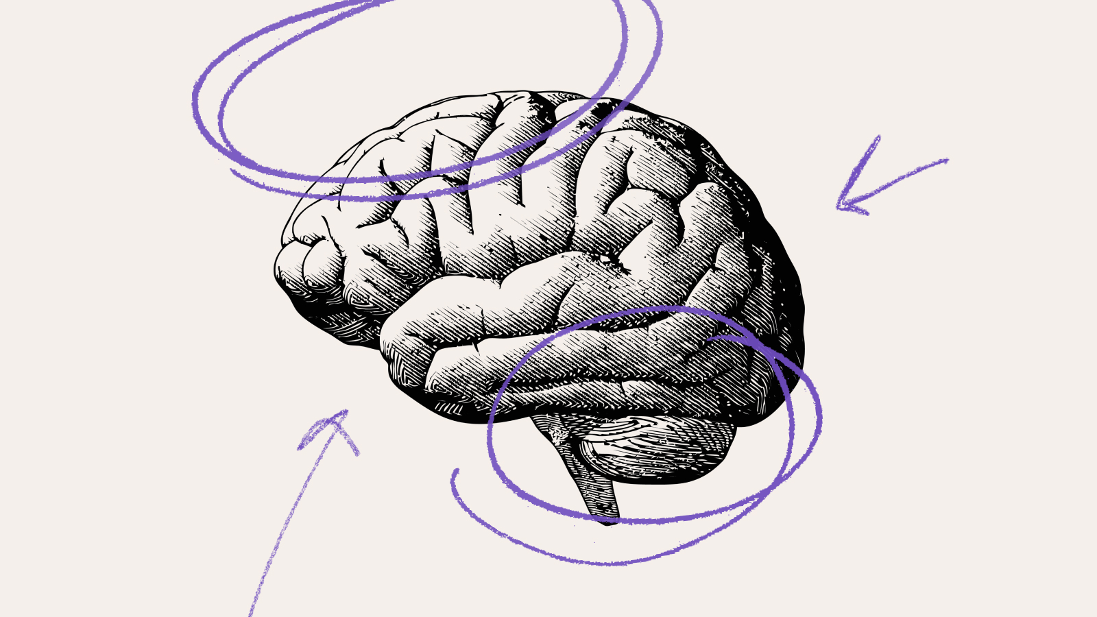
neuroscience
The “dystopian” biotech imagined in these novels is now changing real lives for the better.
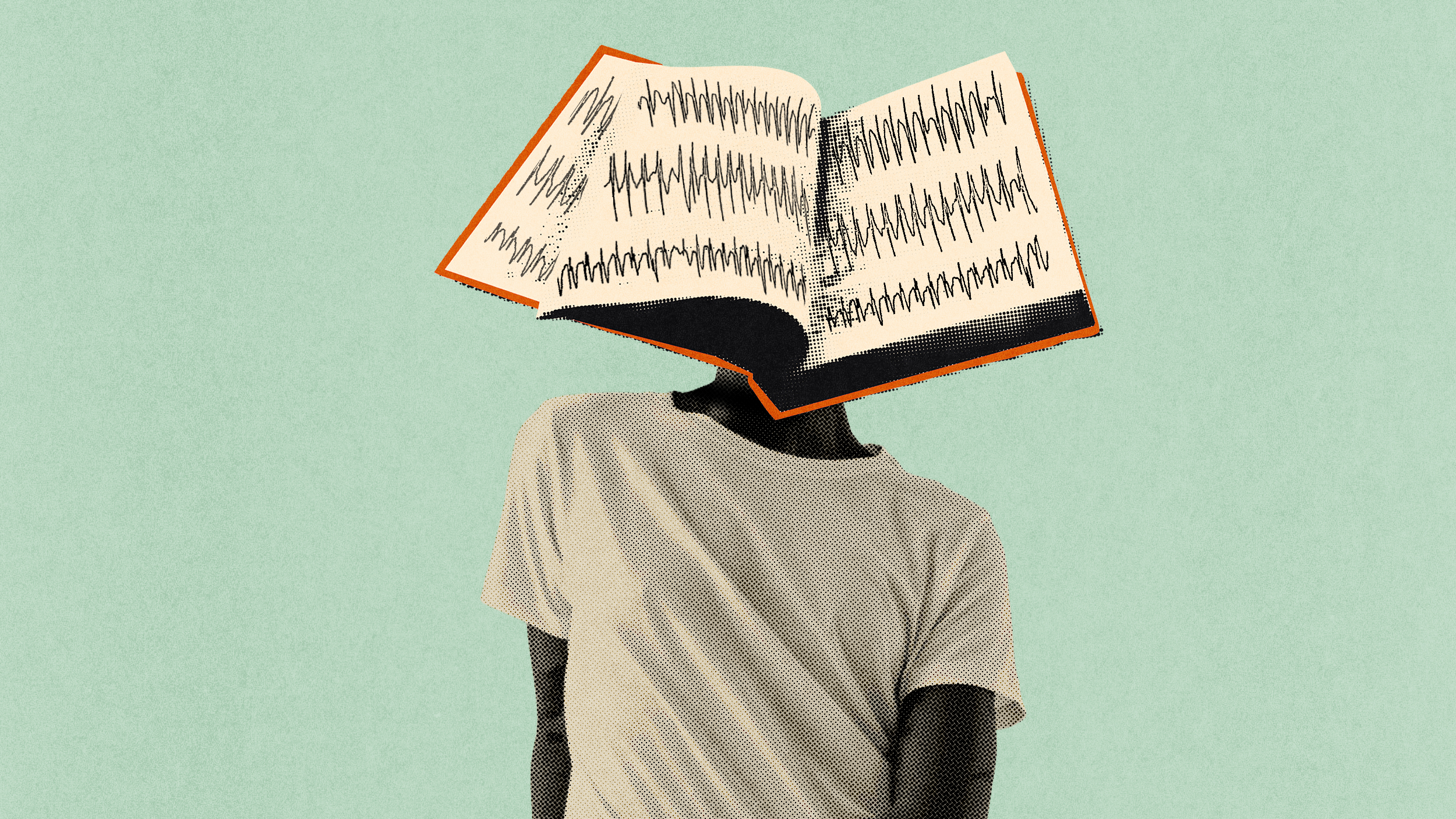
Books don’t just stimulate the mind — they trigger physiological changes throughout the body.
Moltbook is a social media site built for conversation — but not for humans.

Many top performers start behind — and overtake the early leaders later.
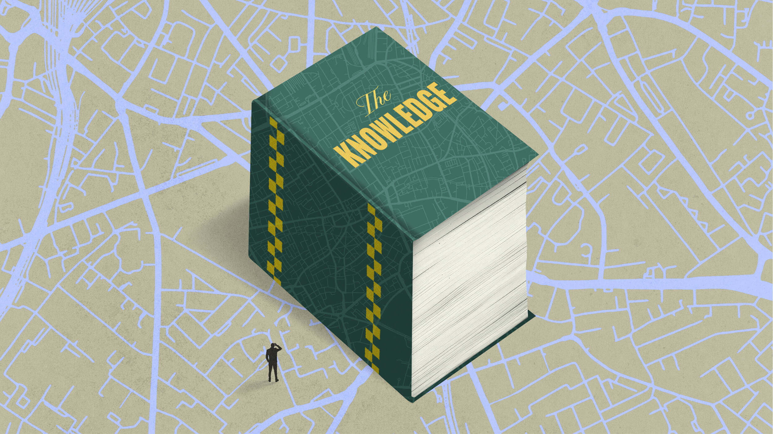
One of the toughest vocational exams in the world requires candidates to memorize 25,000 streets in an area five times the size of Manhattan.

Elite athletes train their “quiet eye.” What happens if the rest of us do the same?

The Stoic philosopher argued that most of life is outside our control — but the little we do control defines who we are.

Researchers built a model that behaves like a brain. Without being trained on neural data, the model produced a peculiar signal — one that was later discovered in actual brain activity.

These cultural lies make normal struggle feel like failure. A habit of experimentation makes it feel like progress.

Neuroscience isn’t dissolving philosophy’s hardest problems — it’s forcing us to rethink where they live.

By tracking brain activity as primates move freely in the wild, neuroethology could reshape what we think we know about our own minds.

Big Think and the John Templeton Foundation gathered scientists, artists, and storytellers in Los Angeles to explore the power of awe.

Rituals serve psychological functions that go far beyond mere habit or tradition.
The technology might be much closer than you’d think.

Metacognition — the ability to think about your thinking — can help you learn faster and make better decisions.
In this excerpt from “Playful,” Cas Holman surveys the research that brought the neuroscience of play into the mainstream.

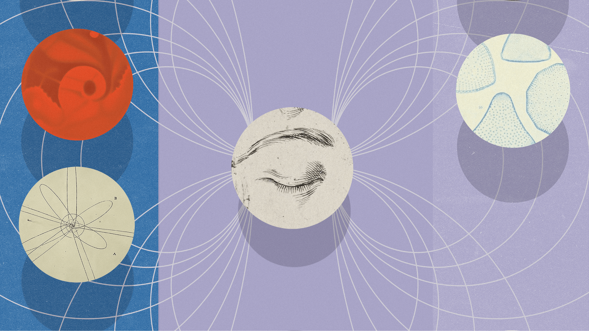
In this excerpt from “One Hand Clapping,” Nikolay Kukushkin makes the case that neurons reveal how memory, meaning, and even consciousness emerge from the same biological roots in humans, sea slugs, and beyond.
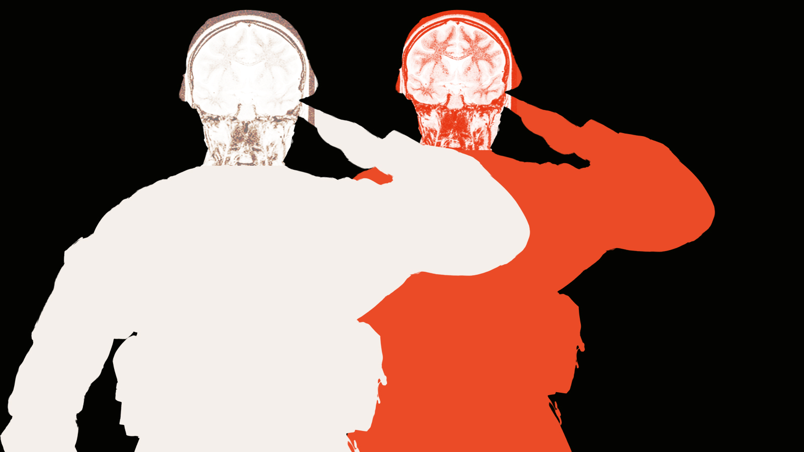
In “Warhead,” neuroscientist and national security adviser Nicholas Wright explains how the brain navigates warfare and why it is our ultimate weapon (and instrument for peace).

Getting drunk might be bad for you but good for us.
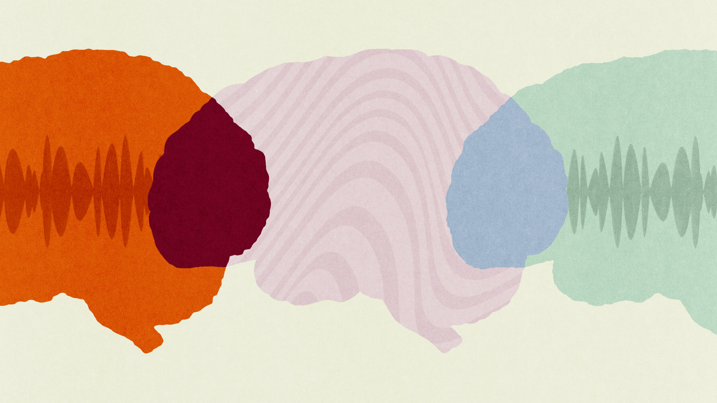
You may actually be on the same wavelength.

Life’s “in-between” stages pack unique cognitive benefits — if you know how to tap into them.
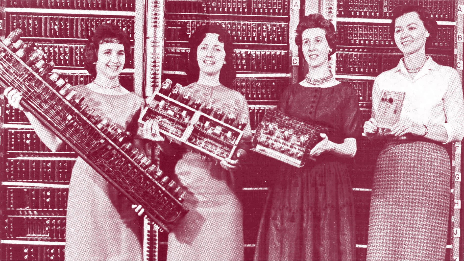
The incredible story of how the US Army began the march toward generative AI in 1943 — and what it means for your business today.
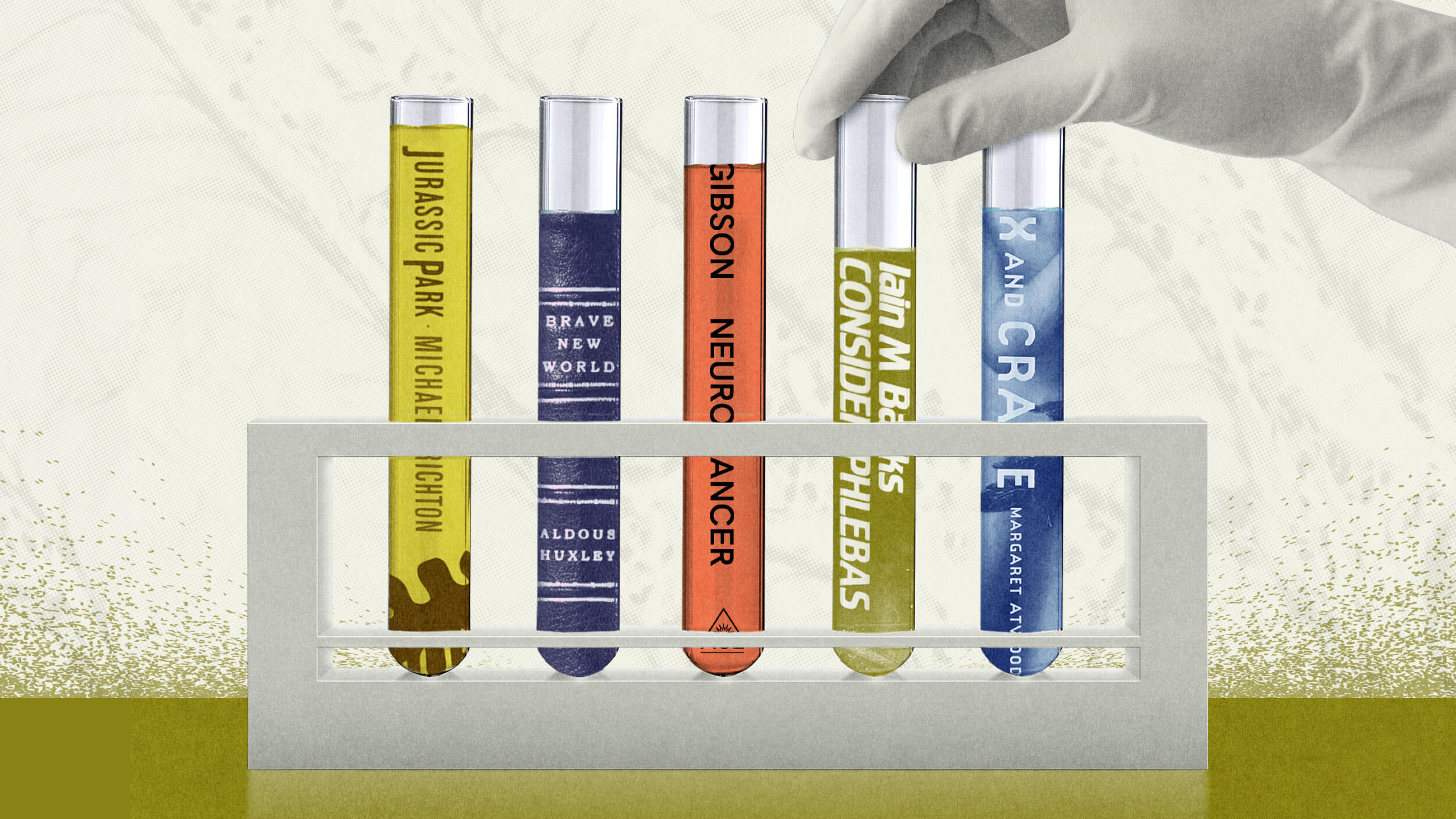
Neuroscientist Rachel Barr shares her favorite books on the brain and how they shaped her approach to the field.

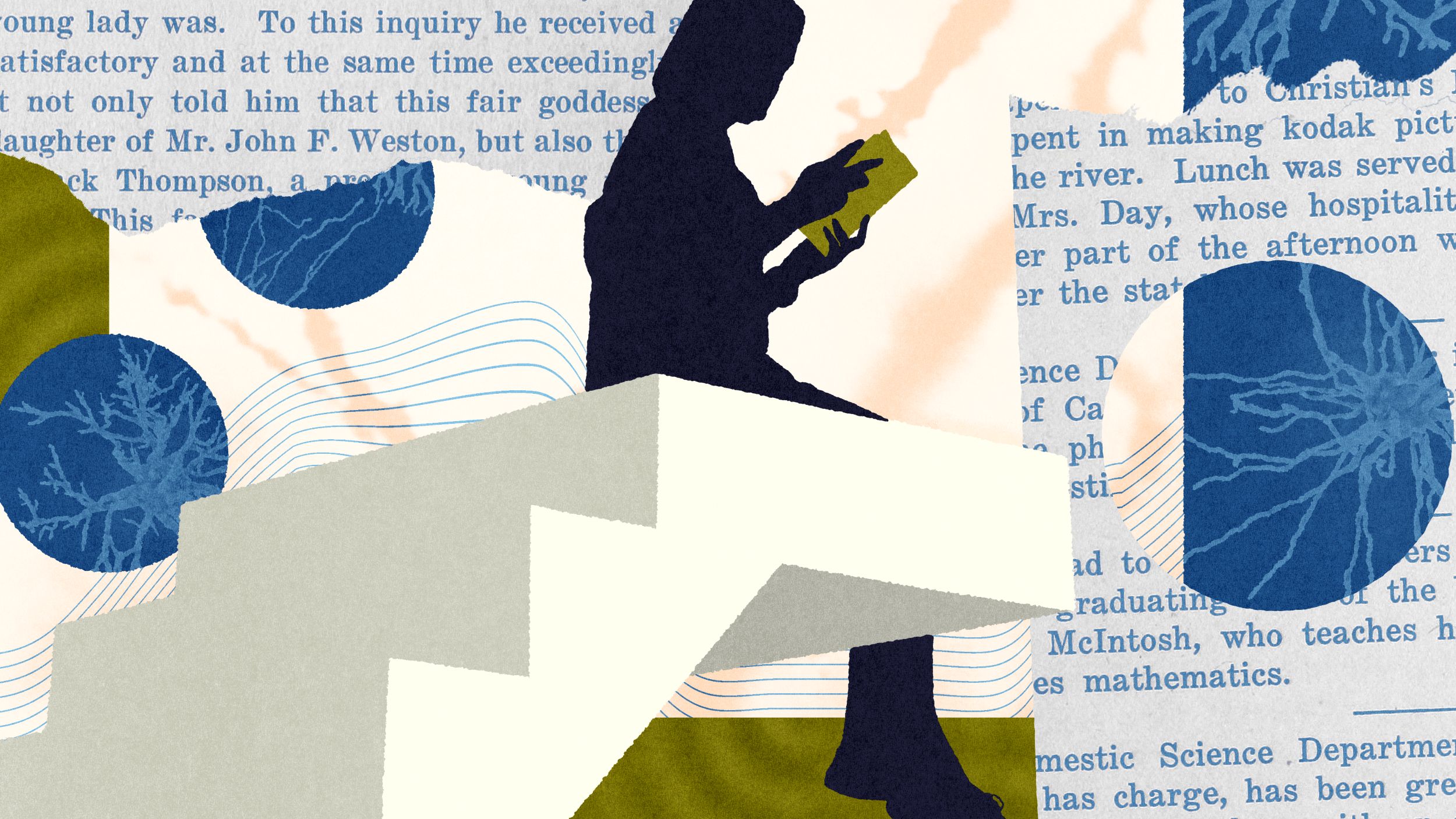
Despite the claims of speed reading apps and programs, you actually have to read the book if you want to learn.

Strengthen your focus like a muscle.
A dialogue with Angus Fletcher — author of the bestseller “Primal Intelligence” — exploring the unique engines of human progress.

A conversation with neuroscientist Erik Hoel about the future of consciousness research.
A conversation with Annaka Harris on shared perception, experimental science, and why our intuition about consciousness is wrong.

“Ordinary dreams are, perhaps, the clearest articulation of what it is like to be.”

The overlooked reason why “AI consciousness” isn’t coming anytime soon.