Transparent Brain Imaging Will Accelerate Research 10 to 100 Times
The world of neuroscience is abuzz with the news that a new technique has been developed to study brain anatomy in mice. By removing the brain and treating it with chemicals, researchers are able to obtain a transparent view.
This advance was made by the bioengineering lab of Dr. Karl Deisseroth at Stanford and reported in the journal Nature yesterday. “Obtaining high-resolution information from a complex system, while maintaining the global perspective needed to understand system function, represents a key challenge in biology,” the scientists wrote.
Their answer to this challenge is called CLARITY, which uses chemicals to transform intact brain tissue into a form that is optically “transparent and macromolecule-permeable.”
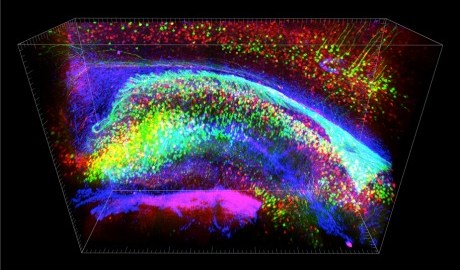
To illustrate this breakthrough, Dr. Deisseroth’s team released two videos. One shows “a flythrough” of a mouse brain using a fluorescent imaging technique. The second shows a 3D view of a mouse brain’s memory hub, or hippocampus.
As the scientists note, existing methods require making hundreds of thin slices to the brain, and most crucially, hinders scientists’ ability to analyze intact components in relation to each other.
So how significant is this development? “It’s exactly the technique everyone’s been waiting for,” Terry Sejnowski of the Salk Institute told the Associated Press, estimating it will speed up brain anatomy research “by 10 to 100 times.”
Watch the CLARITY demo videos here:
Image courtesy of Shutterstock