The end of slicing?

Have you ever sliced up a human brain?
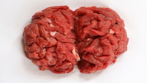
I’ll be honest: I’ve only done it once. I don’t remember much about it–it was a long time ago. But I recall that the consistency and feeling of the brain through my rubber gloves was not at all what I expected (though I could not, for the life of me, tell you what it is that I had expected). Looking back, I think of Jello combined with a skewed breadcrumb-to-ground-meat ratio meatloaf–but even that doesn’t give the tactile interaction justice. It’s firm yet delicate. The color of cat vomit thanks to its Formulin bath. And it’s not quite squishy, though you think that it might be. Honestly, it’s hard to describe. In many ways, the human brain tissue, post-mortem, seems almost other-worldly once it encounters your fingertips. (Though, how much of that is the idea that you’re touching human brain versus it actually being human brain is not something I can ever really know). Regardless, it doesn’t seem like something you should necessarily be touching. And it definitely does not seem like something that you put through the same kind of slicer you might find at your local supermarket deli counter.
But that’s basically what happens. Though, again, I only did it once. My memory may fail me a bit. You don’t let amateurs do this kind of work. Good brains are hard to come by. One wrong move and you lose a lot of valuable data.
After a recent presentation at a large neuroscience conference, I’ve learned that it’s entirely possible, that such brain slicing may soon be a thing of the past. When I spoke with Nora Volkow, the Director of the National Institute on Drug Abuse (NIDA), earlier this week at Neuroscience 2012, she told me about a new technology that has her very excited. It’s called CLARITY — a new project by Stanford University bioengineer, Karl Deisseroth. And she believes it is going to transform the way we do pathology.
CLARITY stands for clear, Lipid-exchanged, Anatomically Rigid, Imaging/immunostaining compatible, Tissue hYdrogel. I know it sounds like a mouthful. But, basically, Deisseroth has figured out a way to do high-throughput whole fixed brain analysis at the cellular level without sectioning or reconstruction. Simply put: lots of staining and looking and modeling without any slicing.
“It allows you to remove the lipids from the brain, which are opaque, and then look at the cells at a 3-dimensional resolution at the microscopic level,” Volkow told me. “So you can see, in a post-mortem brain without having to slice it up, the 3-dimensional structure of specific neurons. You can label those cells with antibodies, you can wash it out and you can create new models using the computer.”
I replied with the first thing that came to mind, the feeling of that not-quite-squishy brain still with me. “That’s really sexy.”
“It’s incredible. It’s so beautiful,” she says. “I was looking at these images and I was just in ecstasy. Not just because of the scientific implications but because of the beauty of seeing these hippocampal cells, and their projections. Being able to get right in there and see–it was amazing.”
Currently, CLARITY is only looking at mouse brains. But it’s not hard to imagine that the technology can be applied to human brains in the near future. And Volkow is looking forward to that.
“It’s going to revolutionize the way we do post-mortem histological studies completely,” she says. “The potential for our understanding of pathology, of the brain–it’s amazing.”
Photo Credit: Radim Strojek/Shutterstock.com


